Shop By Category
- -25°C Biomedical Freezer
- -25°C Chest Freezer
- -25°C Upright Freezer
- -40°C Upright Freezer
- -60°C Chest Freezer
- -86°C Ultra Low Temperature Deep Freezer
- -86°C Ultra Low Temperature Freezer
- -86°C Ultra-Low Chest Freezer
- -86°C Ultra-Low Vertical Freezer
- 1200°C Muffle Furnace
- Abbe Refractometer
- Advanced Thermal Cycler
- Aerosol Photometer
- Air Borne Particle Counter
- Air Samplers
- Anaerobic Incubator
- Anaerobic Jar
- Analytical Balance
- Analytical HPLC System
- Analyzers
- Atmosphere Furnace
- Auto Refractometer
- Autoclave Steam Sterilizer
- Autoclave
- Automated Nucleic Acid Extractor
- Automatic Asphalt Tester
- Automatic Chemistry Analyzer
- Automatic Colony Counter
- Automatic Lifting Rotary Evaporator
- Automatic Microtome
- Automatic Slide Stainer
- Automatic Tissue Processors
- Bacti Cinerator Sterilizer
- Ball Mill Machine
- Beam Balance
- Bench Hood
- Bench Top Autoclave
- Benchtop Multi-parameter Water Quality Meter
- Benchtop Ph Meter
- Benchtop Water Activity Meter
- Bilirubinometer
- Biochemical Incubator
- Biochemistry Analyzer
- Biological Indicator Incubator
- Biological Microscope
- Biological Safety Cabinet Class II A2
- Biosafety Cabinets
- Blood Bank Refrigerator
- Blood Card Centrifuge
- Blood Cell Counter
- Blood Coagulation Analyzer
- Blood Collection Monitor
- Blood Donor Chair
- Blood Serum Centrifuge
- Blood Warmer
- Blood Weighing Scale
- Carbon-dioxide Incubator
- Cell Counter
- Cell Washer Centrifuge
- Chemical Resistant Fume Hood
- Chemiluminescence Imaging System
- Chemiluminescence Immunoassay Analyzer
- Chiller
- Chlorophyll Meter
- Coating Thickness Gauge
- COD Thermoreactor
- Colony Counter
- Colorimeter
- Combined Refrigerator And Freezer
- Conductivity Meter
- Confocal Microscope
- Constant Temperature Water Bath
- Cryostat Microtome
- Deep Freezer
- Density Meter
- Dental Vacuum Autoclave
- Diaphragm Vacuum Pump
- Digital Automatic Polarimeter
- Digital Colony Counter
- Digital Hot Plate
- Digital Hot-Plate
- Digital Magnetic Hot Plate Stirrer
- Digital Multi Tester
- Digital Peristaltic Pump
- Disintegration Tester
- Double Beam UV-Vis Spectrophotometer
- Drug Test Chamber
- Dry Bath Incubator
- Dry Block Temperature Calibrator
- Drying Oven
- Ducted Fume Hood
- Ductless Fume Hood
- Egg Incubator
- Electroencephalogram Machine
- Electrolyte Analyzer
- Electronic Pipette Filler
- Electrophoresis Power Supply
- Environmental Test Chamber
- Filter Press
- Fixed Volume Pipette
- Flake Ice Maker
- Flame Photometer
- Flow Cytometer
- Fluorescence Quantitative PCR System
- Forced Air Vacuum Drying Oven
- Formaldehyde Gas Detector
- Freeze Dryer
- FTIR Spectrometer
- Fume Hoods
- Gas Analyzer
- Gas Chromatography
- Gas Chromatography Mass Spectrometry (GC-MS)
- Gas Detectors
- Gel Card Incubator Centrifuge
- Gel Electrophoresis 2D
- Gel Electrophoresis System
- Gel-Documentation Imaging System
- Gel-Documentation System
- Glass Bead Sterilizer
- Glucometer
- Glucose Meter
- Gradient Thermal Cycler
- Graphite Hot Plate
- Halogen Moisture Analyzer
- Handheld XRF Spectrometer
- Hematocrit Centrifuge
- Hematology Analyzer
- Hemoglobin Analyzer
- High Pressure Portable Autoclave
- High Shear Homogenizer
- High Speed Centrifuge
- High Speed Refrigerated Centrifuge
- High-Flow Peristaltic Pump
- High-Performance Liquid Chromatography (HPLC)
- Homogenizer
- Horizontal Autoclave
- Horizontal Gel Electrophoresis
- Horizontal Laminar Flow Cabinet
- Hot Air Sterilizer
- Hot-Plate Magnetic Stirrer
- Ice Maker
- ICP Mass Spectrometer
- Imaging System
- Immunohistochemistry stainer
- Incubator
- Incubator Shaker
- Inverted Biological Microscope
- Inverted Fluorescence Microscope
- IR Carbon Sulphur Analyzer
- Kinematic Viscometer
- Kjeldahl Digestor
- Kjeldahl Nitrogen Analyzer
- Kjeldahl Systems
- Lab Dehumidifier
- Lab Spray Dryer
- Laboratory Balances
- Laboratory Centrifuge
- Laboratory Clean Bench
- Laboratory Digesters
- Laboratory Freezers
- Laboratory Furnace
- Laboratory Glassware Washer
- Laboratory Mixers
- Laboratory Oven
- Laboratory Refrigerators
- Laboratory Shaking Table
- Laminar Air Flow Sterilizer
- Laminar Flow Cabinet
- Large Capacity High Speed Refrigerated Centrifuge
- Large Capacity Low Speed Refrigerated Centrifuge
- Large Scale Freeze Dryer
- Laser Diffraction Particle Size Analyzer
- Laser Dust Particle Counter
- Lighting Incubator
- Linear Decolorizing Shaker
- Low Speed Refrigerated Centrifuge
- Low Speed Table Top Centrifuge
- Magnetic Sensor Analytical Balance
- Magnetic Stirrer
- Manual Lifting Rotary Evaporator
- Manual Polarimeter
- Medical Refrigerator
- Melting Point Apparatus
- Melting Point Meter
- Micro Spectrophotometer
- Microbial Air Sampler
- Microbial Identification System
- Micropipettes
- Microplate Centrifuge
- Microplate Incubator
- Microplate Reader
- Microplate Reader And Washer
- Microplate Washer
- Microscope
- Microscope Slide Cabinet
- Microtome
- Microwave Digesters
- Mini Laminar Flow Cabinet
- Mini Shaker
- MIRI (Mini Incubator for Research and Innovation)
- Mixers And Vortexer
- Moisture Analyzer
- Mold Incubator
- Multi Tube Vortex Mixer
- Multi-channel Hotplate Magnetic Stirrer
- Multi-Channel Pipette
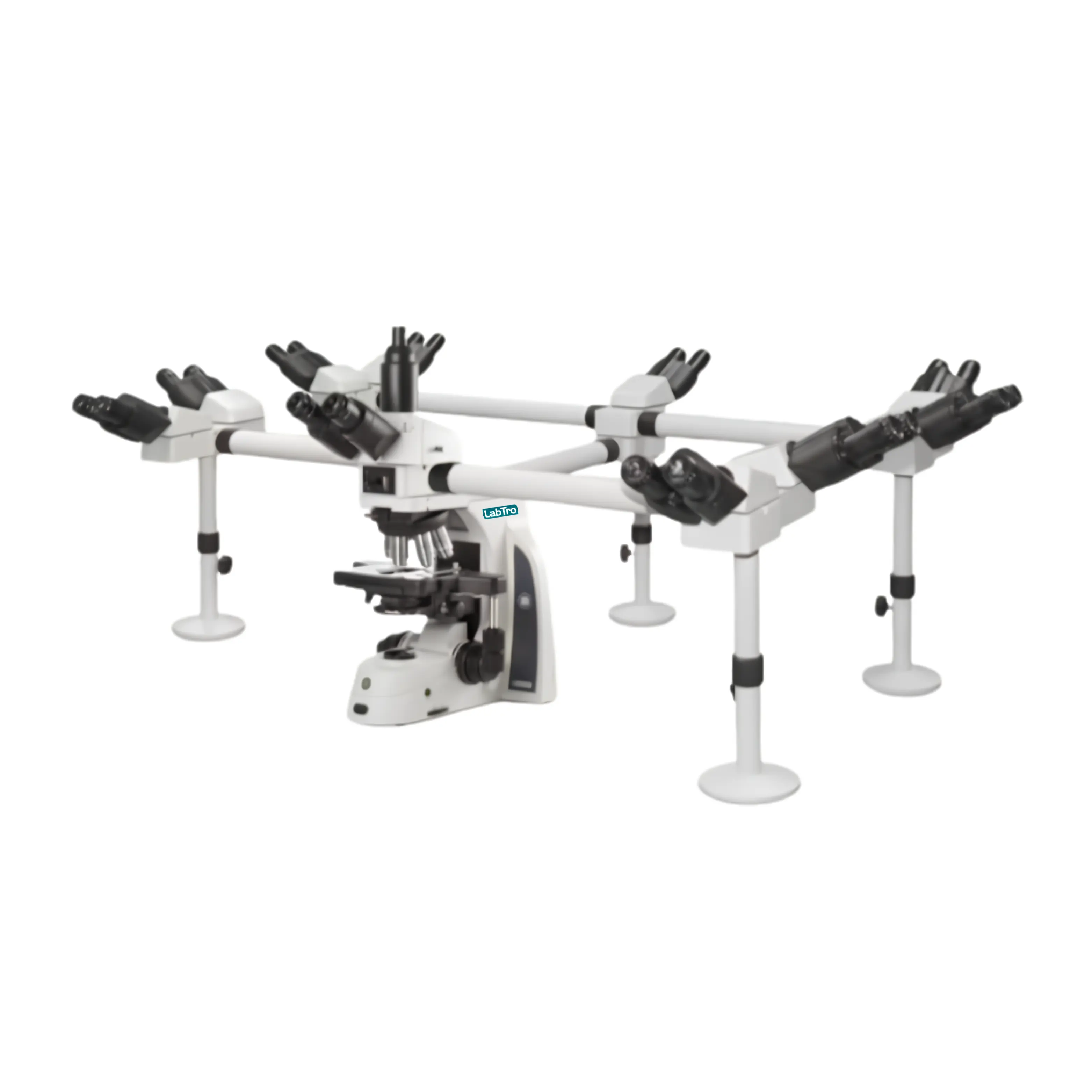
- Multi-Viewing Microscope
- Nano Spectrophotometer
- Nitrite Nitrogen Analyzer
- Nucleic Acid Analyzer
- Nucleic Acid Extractor
- Nucleic Acid Extractor System
- Orbital Shaker
- Overhead Stirrer
- Oxygen Concentrator
- Particle Counter
- Particle Size Analyzer
- PCR System
- Perchloric Acid Fume Hood
- Peristaltic Pump
- Petroleum Testing Equipment
- PH Meter
- Pharmaceutical Freeze Dryer
- Pharmacy Refrigerator
- Photometer
- Planetary Ball Mill
- Plasma Thawing System
- Polarimeter
- Polarizing Microscope
- Portable Dissolved Oxygen Meter
- Portable Freezer
- Portable Gas Monitor
- Portable Ion Meter
- Portable PH Meter
- Portable Refractometer
- Portable Water Activity Meter
- Potentiometric Titrator
- Precision Balance
- Pulse Vacuum Autoclave
- Pulse Vacuum Sterilizer
- Pumps
- Real Time PCR
- Recirculating Chiller
- Refractometer
- Refrigerated Centrifuge
- Refrigerated Water Bath
- Rocking Shaker
- Roller Compactor
- Roller Shaker
- Rotary Evaporator
- Rotary Microtome
- Rotary Mixer
- Rotation Mixer
- Rotational Viscometer
- Safety Valve Test Bench
- Seal Integrity Tester
- Semi Automatic Microtome
- Shakers
- Shaking Incubator
- Shaking Water Bath
- Single Beam UV-VIS Spectrophotometer
- Single Beam VIS Spectrophotometer
- Slide Warmer
- Soxhlet Apparatus
- SPAD Chlorophyll Meter
- Spectrometers
- Spectrophotometer
- Spin Tissue Processor
- Spirometer
- Stability Test Chamber
- Stainers
- Stainless Steel Fume Hood
- Stainless Steel Hot Plate
- Stainless Steel Water Bath
- Stainless Steel Water Distiller
- Steam Sterilizer
- Stereo Microscope
- Sterilizer
- Sterilizer
- Stirrers
- Strong Illumination Test Chamber
- Table Top Autoclave
- Tablet Hardness Testers
- Temperature And Humidity Test Chamber
- Temperature Calibrator
- Test Chamber
- Thermal Cycler
- Thermostatic Bath
- Thermostatic Incubator
- Thermostatic Refractometer
- Thermostatic Water Bath
- Thickness Gauge
- Thin Layer Chromatography
- Tissue Embedding System
- Tissue Water Bath
- Titrator
- Total Organic Carbon Analyzer
- Transparent Water Bath
- Tube Roller Mixer
- Tube Sealer
- Turbidity Meter
- Ultrapure Water System
- Ultrasonic Cleaner
- Ultrasonic Homogenizer
- Urine Analyzer
- Vacuum Drying Oven
- Vacuum Freeze Dryer
- Vacuum Leakage Tester
- Vacuum Muffle Furnace
- Variable Speed Peristaltic Pump
- Variable Volume Pipette
- Vein Locator
- Vertical Autoclave
- Vertical Clean Bench
- Vertical Gel Electrophoresis System
- Vertical Laminar Flow Cabinet
- Vibrating Microtome
- Vibration Meter
- Viscometer
- Volumetric Karl Fischer Titrator
- Vortex Mixer
- Water Bath
- Water Circulating Vacuum Pump
- Water Distillers
- Water Purification System
- Water Quality Analyzer
- Water Quality Meters
- Water Testing Equipment
- Water-Jacketed CO2 Incubator
- X-ray Fluorescence Spectrometer
- Xray Fluorescence (XRF) Spectrometer